Information for patients
If you are a patient reading this and have a concern about an MRI scan you are scheduled to attend, we strongly recommend you contact the site where your scan is due to take place, you may also wish to refer to our ‘Information for Patients’ section. Please note local variations to the policies detailed on this website may apply, therefore please contact the hospital where your appointment is scheduled for clarification.Disclaimer (MUST READ)
The MRI safety information contained within this webpage is intended for use by staff from NHS Greater Glasgow and Clyde (GGC) and associated health boards, namely: NHS Ayrshire and Arran, NHS Borders, NHS Dumfries & Galloway, NHS Forth Valley, NHS Golden Jubilee, and NHS Lanarkshire. Only staff from these health boards are approved to use this information and local variations to the policies detailed may apply. Non-approved users i.e. patients and staff from health boards other than those listed above, or staff from private medical organisations use this information at their own risk. We, NHS GGC, accept no responsibility for patient injury or adverse outcomes that may occur as a consequence of the information contained herein. If you have any questions regarding this disclaimer, please contact the NHS GGC MRI physics team on: ggc.MRSafetyExpert@nhs.scot.NHS GGC MRI safety policy for scanning patients with Cerebral vascular embolisation/aneurysm coils and embolisation devices
Brief summary of known devices
Cerebral vascular embolisation/aneurysm coils and embolisation devices.
Device use
Embolisation/aneurysm coils and devices are used to provide vascular occlusion, including the treatment for aneurysms. The device is inserted into the aneurysm or vessel, reducing blood flow and promoting clotting. Multiple coils are typically used for aneurysms and large vessel occlusion but have a range of uses including applications where the coil may be uncoiled or elongated.
Liquid embolic agents are rapidly solidifying agents that are also used to provide vascular occlusion, including into an aneurysm or AVM lumen.
Radioembolisation microspheres are glass or resin spheres containing Yttrium-90 that can be used to embolise unresectable tumours.
Flow-diverting stents are used to divert the flow of blood away from the aneurysm. They are often used in combination with embolisation coils and the procedure is often referred to as “stent-assisted coiling”.
WEB aneurysm embolisation devices are used to provide vascular occlusion, including the treatment for aneurysms and other neurovascular abnormalities such as arteriovenous fistulae (AVF). The device is inserted into the aneurysm or vessel, reducing blood flow and promoting clotting.
Must read: What this policy does not cover / notable exceptions
This GISP does not cover coils implanted outside the cerebral vascular area.
This GISP does not cover aneurysm clips, nor situations where any non-embolisation implants are placed in close proximity to embolisation implants. Situations where the catheter is broken and retained are also excluded from this GISP and must be investigated on a case-by-case basis.
If a patient reports that their coil was inserted as part of a clinical trial or research study, these must be investigated on a case-by-case basis.
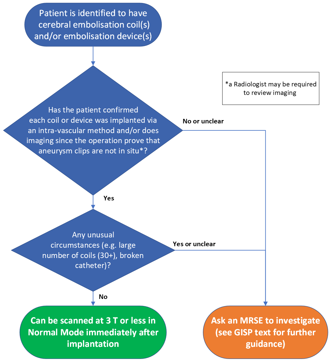
Embolisation coiling can be very inconsistent in the approaches used, if there are any unusual circumstances these should be considered outside this GISP. These circumstances can be identified at the time of screening or from previous imaging and may include a large number of coils (30+) in close proximity. It is at the radiographer’s professional discretion what is deemed an “unusual circumstance”.
Must read: What the policy covers
This policy covers 1.5 and 3T MRI scanning of patients with cerebral vascular embolisation coils, WEB aneurysm embolisation devices, flow-diverting stents, liquid embolic devices and radioembolisation microspheres.
Must read: The MR safety policy
Particular care must be taken to confirm cerebral aneurysm embolisation coils and other embolisation implants are not confused with aneurysm clips. If a referrer or patient reports that a patient has cerebral aneurysm coils or other embolisation implants, the patient must be asked how the procedure or operation was performed (e.g. identify if it was via an intra-vascular procedure or craniotomy). If there is any doubt, a review of prior imaging or clinical notes is recommended to exclude the presence of an aneurysm clip. If an aneurysm clip cannot be ruled out, the make and model of each implant must be identified or a screening x-ray may be required as part of a risk-benefit analysis.
If the implant(s) fall within the remit of the GISP as outlined above, they can undergo an MR examination at 3T or less, in Normal Operating Mode, at any time after coil implantation. This advice is regardless of the value of the spatial gradient of the MRI scanner. Note that this information applies to cases in which the coils are in the area of the transmitted RF. If no clinical need to scan at 3T and 1.5T is available, scan at 1.5T.
For stent-assisted coiling, please also follow the non-coronary vascular stent policy.
Detailed review and risk assessments
The documentation underpinning this policy can be found here:
Embolisation coils and devices GISP documentation
Additional background information and discussion
There will be significant susceptibility artefact from stainless steel embolisation coils that may limit the usefulness of the MR examination if the coils are near the area of interest. For platinum coils that are used more frequently, the susceptibility artefact is greatly reduced.
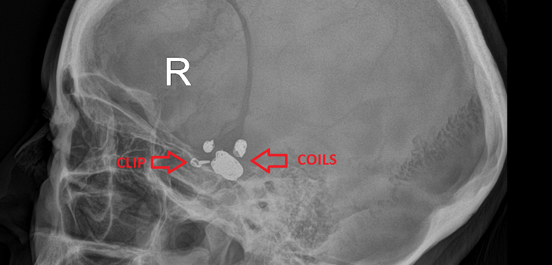
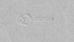
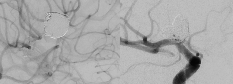
The following images are provided as an example of how an aneurysm clip and embolisation coils may appear in prior imaging. If in any doubt, images should be reviewed by a Consultant Radiologist and further advice can be provided from MR Physics.
There will likely be significant signal loss within a WEB device such that MRI cannot be used to confirm complete thrombus formation within the implant.